Diagnosis and workup
There is no singular test for Mycosis Fungoides or Sézary Syndrome. Accurate diagnosis requires skin biopsy, as well as close correlation between clinical, histopathological, and genotypic results.1,2
A multimodal diagnostic workup is recommended, including history, complete physical exam, and skin biopsy. Blood evaluation and imaging studies may be helpful if clinically indicated.1
History and complete physical exam
- Identify lesion types
- Quantify % BSA affected
- Palpate for lymph node enlargement and visceral masses
- Lab tests, including CBC, metabolic panel, LDH level
Skin biopsy
- Dermatopathology
- lmmunophenotyping for T-cell surface markers
- Molecular analysis to test for clonality
Blood evaluation
- Flow cytometry to assess and quantitate abnormal T cells
- TCR gene rearrangement assessment
Lymph node assessment
- Palpation and/or biopsy, if clinically indicated
- Evaluation of nodal architecture, pathologic assessment
Imaging studies
- Recommended for skin classification ≥T2 (C/A/P CT with contrast or integrated whole-body PET-CT)
- Consider for other stages, if clinically indicated
The International Society for Cutaneous Lymphoma (ISCL) has proposed a diagnostic algorithm for early Mycosis Fungoides based on clinical, histological, immunophenotypical, and molecular criteria. Using this algorithm, a total of four points is required for a diagnosis of early Mycosis Fungoides.3,4
| ISCL Algorithm for the Diagnosis of Early Mycosis Fungoides | Major | Minor |
|---|---|---|
| Criteria | 2 points | 1 point |
| Clinical | Any 2 | Any 1 |
|
||
| Histopathologic | Both | Either |
|
||
| Molecular/biologic |
Present | |
|
||
| lmmunopathologic | Any 1 | |
|

Assessing skin involvement
Punch biopsy
Biopsy of suspicious skin lesions is essential for diagnosis. Multiple biopsies may be necessary to capture the pathologic variability of disease at diagnosis.5
Punch biopsy–preferred for MF and SS


A circular blade (6-mm diameter preferred) is rotated through several layers of the skin to produce a cylindrical core tissue.3
Considerations for skin biopsy technique5
Guidance for skin biopsy when Mycosis Fungoides is suspected was developed by an international panel of dermatologists, oncologists, hematologists and dermatopathologists:
- Punch biopsy, 6-mm needle size is recommended
- Perform deeper-punch biopsies if there is clinical suspicion of adnexotropic/folliculotropic involvement
- Consider taking biopsies from several lesions if lesions appear variable
- Discontinuation of topical steroids ≥2 weeks before biopsy is recommended, because topical medications can affect biopsy results
- Light exposure may also affect results; patients should be advised to avoid sunbathing
If biopsy is inconclusive, monitor lesions for changes or progression.5
Multiple biopsies may be needed
- Skin biopsies can be inconclusive—one study found that the first biopsy is diagnostic only 20% to 25% of the time6
- Biopsy results may vary over time and between multiple sites in the same patient5
- Often there is a substantial delay in histopathologic diagnosis—a recent PROCLIPI (an international registry of mycosis fungoides PROspective Cutaneous Lymphoma International Prognostic Index) study reported a median delay of 3 years from onset to definitive diagnosis2
Considerations in assessing skin
Consider discontinuing topical steroids and other skin-directed therapy 2 weeks prior to obtaining skin biopsies in order to maximize the likelihood of accurate diagnosis.1,5
Consider a comprehensive morphological and molecular review of a skin biopsy and a peripheral blood sample to rule out a diagnosis of CTCL before introducing a biologic treatment.7,8
Histopathologic findings of Mycosis Fungoides, even in cases showing classic features, need to be correlated with clinical presentation in order to reach a definitive diagnosis.5
Consider T-cell receptor (TCR) gene rearrangement analysis (either polymerase chain reaction [PCR] or next-generation sequencing). Rebiopsy skin if1:
- Pathology findings are non-diagnostic and/or discordant with the clinical presentation (eg, if negative but clinical suspicion of CTCL is high)
- If large-cell transformation (LCT) is suspected clinically
Consider flow cytometry and molecular analysis to detect clonal TCR gene rearrangements or other assessment of clonality if Mycosis Fungoides or Sézary Syndrome is suspected. It is expected that patients with high blood tumor burden will have identical clones in blood and skin.9
- Immunohistochemical evaluation of the skin plays an important diagnostic role. T-cell markers, including CD3, CD4, CD5, CD7, CD8, and CD20, can help confirm diagnosis5
- In cases where Sézary Syndrome is suspected but skin biopsies are inconclusive, peripheral blood flow cytometry should be performed for more definitive diagnosis
Multiple biopsies taken over time may help confirm an inconclusive diagnosis.5
Rebiopsy skin if large-cell transformation or folliculotropic Mycosis Fungoides is suspected, or if aggressive clinical behavior.1
Evaluation of skin biopsy by pathologist with expertise in CTCL is recommended.1

Challenges in diagnosis
In a SEER population data analysis, less than 15,000 new cases of CTCL were identified in a 19-year period.8 This rarity can lead to diagnostic delays (eg, being treated for a “rash” for many years prior to a diagnosis of Mycosis Fungoides).11
Heterogeneity of presentation
Skin symptoms may look different from one patient to another. Classic Mycosis Fungoides presents with patches, plaques, and tumors. However, less common variants may present differently, such as with lesions that are hypo- or hyperpigmented, or with other rare variants such as poikilodermatous Mycosis Fungoides. Further, lesion types may present in combination (eg, ulcerated tumor and flat patches).12-15
Mimics more common skin diseases12
Mycosis Fungoides and Sézary Syndrome tend to resemble other inflammatory skin diseases, such as psoriasis, eczema, or atopic dermatitis.14 This can contribute to delays in diagnosis.2,16,17
Accurate diagnosis is important because emerging evidence suggests the use of certain biologic treatments used for inflammatory skin conditions (eg, psoriasis) may result in more rapid disease progression.2,7,8,18,19 Previous treatments may also confound diagnosis, such as topical steroids and skin-directed therapy. Even sun exposure may result in false-negative skin biopsy results.5,11
Mycosis Fungoides and Sézary Syndrome vs other skin conditions
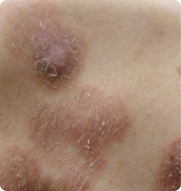
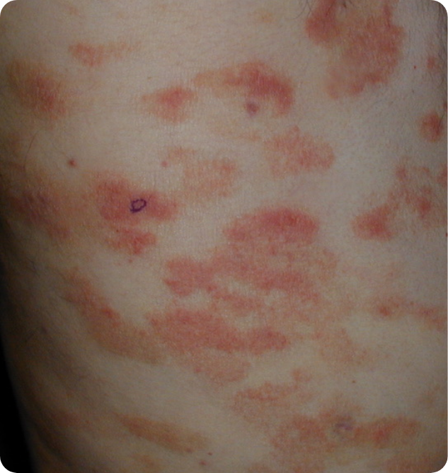
Mycosis Fungoides plaquea
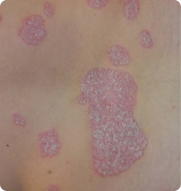
Psoriasis plaqueb
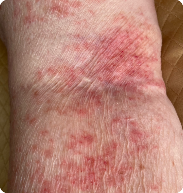
Mycosis Fungoides patcha

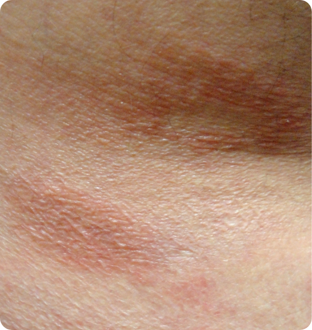
Lichenified eczemac
Sézary – exfoliative erythrodermaa
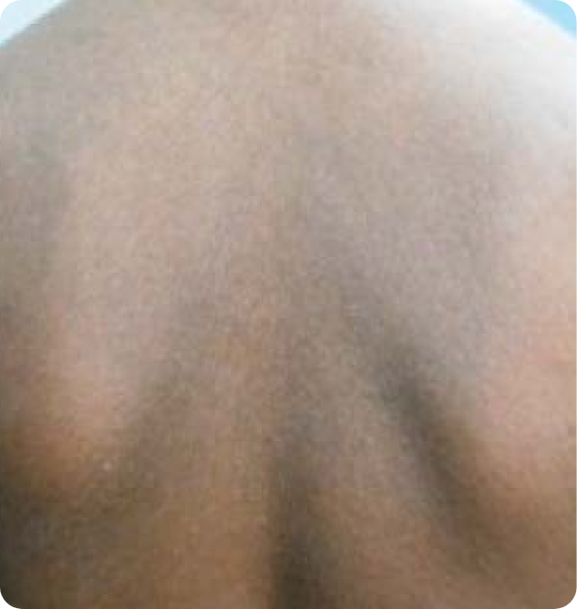
Nonspecific erythrodermad
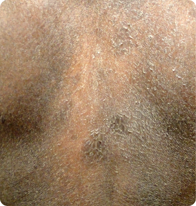
Mycosis Fungoidese
Drug rasha
- Images are intended to be an example. Actual presentation can vary depending on individual patient factors.
- aImages provided courtesy of Dr. Oleg Akilov, University of Pittsburgh.
- bImage used with permission from Dove Medical Press.
- cImage used with permission from J Clin Med.
- dImage used with permission from Ind J of Derm, Ven and Lep.
- eImage provided courtesy of Dr. Joan Guitart, Northwestern University.
Skin symptoms may be more challenging to identify in patients with darker skin
- Patients with skin of color (SOC) are more likely to present with hypo- or hyperpigmentation, silver hue, and lichenification2
- Non-SOC patients more commonly present with erythema and poikiloderma2
Test your knowledge
Question 1 of 4
Below are two photos of erythroderma: one is associated with Sézary Syndrome, and one is not. Which is Sézary Syndrome?
![]() Correct
Correct
As you can see, skin symptoms of Sézary Syndrome can mimic other inflammatory skin conditions.12 This is a major challenge for clinicians and pathologists that can lead to misdiagnosis, and to the use of potentially harmful immunosuppressive agents in these patients.2,7
NCCN Guidelines recommend the use of flow cytometry for evaluation of CTCL stage.1
![]() Incorrect
Incorrect
As you can see, skin symptoms of Sézary Syndrome can mimic other inflammatory skin conditions.12 This is a major challenge for clinicians and pathologists that can lead to misdiagnosis, and to the use of potentially harmful immunosuppressive agents in these patients.2,7
NCCN Guidelines recommend the use of flow cytometry for evaluation of CTCL stage.1
Explore videos for Mycosis Fungoides and Sézary Syndrome
Challenges in diagnosis
Learn more about
TNMB staging
References: 1. Referenced with permission from the NCCN Clinical Practice Guidelines in Oncology (NCCN Guidelines®) for Cutaneous Lymphomas V.2.2026. © National Comprehensive Cancer Network, Inc. 2026. All rights reserved. Accessed March 3, 2026. To view the most recent and complete version of the guideline, go online to NCCN.org. NCCN makes no warranties of any kind whatsoever regarding their content, use or application and disclaims any responsibility for their application or use in any way. 2. Scarisbrick JJ, Quaglino P, Prince HM, et al. The PROCLIPI international registry of early-stage mycosis fungoides identifies substantial diagnostic delay in most patients. Br J Dermatol. 2019;181(2):231-232. 3. Olsen EA, Vonderheid E, Pimpinelli N, et al. Revisions to the staging and classification of mycosis fungoides and Sézary syndrome: a proposal of the International Society for Cutaneous Lymphomas (ISCL) and the cutaneous lymphoma task force of the European Organization of Research and Treatment of Cancer (EORTC). Blood. 2007;110:1713-1722. 4. Miyaki T. Diagnosis of early mycosis fungoides. Diagnostics (Basel). 2021;11(9):1721. 5. Hodak E, Geskin L, Guenova E, et al. Real-life barriers to diagnosis of early mycosis fungoides: an international expert panel discussion. Am J Clin Dermatol. 2023;4(1):5-14. 6. Skov AG, Gniadecki R. Delay in the histopathologic diagnosis of mycosis fungoides. Acta Derm Venereol. 2015;95:472-475. 7. Martinez-Escala ME, Posligua AL, Wickless H, et al. Progression of cutaneous lymphoma after anti-tumor necrosis factor alpha therapy. J Am Dermatol. 2018;78:1068-1076. 8. Kolkowsi K, Sokolowska-Wojdylo M. Safety and danger of biologic treatments in psoriasis in context of cutaneous T-cell lymphoma (CTCL). Adv Dermatol Allergol. 2021;38(6):953-960. 9. Olsen EA, Whittaker S, Willemeze R, et al. Primary cutaneous lymphoma: recommendations for clinical trial design and staging update from the ISCL, USCLC, and EORTC. Blood. 2022;140(5):419-437. 10. Cai ZR, Chen ML, Weinstock MA, et al. Incidence trends of primary cutaneous T-cell lymphoma in the US from 2000 to 2018: a SEER population data analysis. Jama Oncol. 2022;8(11):1691-1692. 11. Lucas AS, Ciccolini K. Nursing best practice referral algorithm for the early detection of mycosis fungoides. J Dermatol Nurse Assoc. 2016;8:109-120. 12. Jawed SI, Myskowski PL, Horwitz S, et al. Primary cutaneous T-cell lymphoma (mycosis fungoides and Sézary syndrome) Part I. Diagnosis: Clinical and histopathologic features and new molecular and biologic markers. J Am Acad Dermatol. 2014;70(2):e1-e16. 13. Cerroni L. Mycosis fungoides—clinical and histopathologic features, differential diagnosis, and treatment. Semin Cutan Med Surg. 2018;37(1):2-10. 14. Hoppe RT, Kim YH. Clinical manifestations, pathologic features, and diagnosis of mycosis fungoides. UpToDate. https://www.uptodate.com/contents/clinical-manifestations-pathologic-features-and-diagnosis-of-mycosis-fungoides. Updated September 19, 2024. Accessed December 18, 2024. 15. Virmani P, Myskowski PL, Pulitzer M. Unusual variants of mycosis fungoides. Dian Histopath (Oxf). 2016;21(22):142-151. 16. Hoppe RT, Kim YH. Clinical manifestations, pathologic features, and diagnosis of mycosis fungoides. UpToDate. https://www.uptodate.com/contents/clinical-manifestations-pathologic-features-and-diagnosis-of-mycosis-fungoides. Updated September 23, 2022. Accessed October 24, 2022. 17. Mangold AR, Thompson AK, Davis MD, et al. Early clinical manifestations of Sézary syndrome: A multicenter retrospective cohort study. J Am Acad Dermatol. 2017;77(4):719-727. 18. Ogilvie C, Jackson R, Leach M, McKay P. Sezary syndrome: diagnosis and management. J R Coll Physicians Edinb. 2012;42(4):317-321. 19. Schaefer L, Comfere N, Sokumbi O. Development of cutaneous T-Cell lymphoma following biologic treatment: A systematic review. Am J Clin Dermatol. 2023;24(2):153-164. 20. Park A, Wong L, Lang A , et al. Cutaneous T-cell lymphoma following dupilumab use: a systematic review. Int J Dermatol. 2023;62(7):862-876.